Sasank Chilamkurthy, Rohit Ghosh, Swetha Tanamala, Mustafa Biviji, Norbert G. Campeau, Vasantha Kumar Venugopal, Vidur Mahajan, Pooja Rao and Prashant Warier
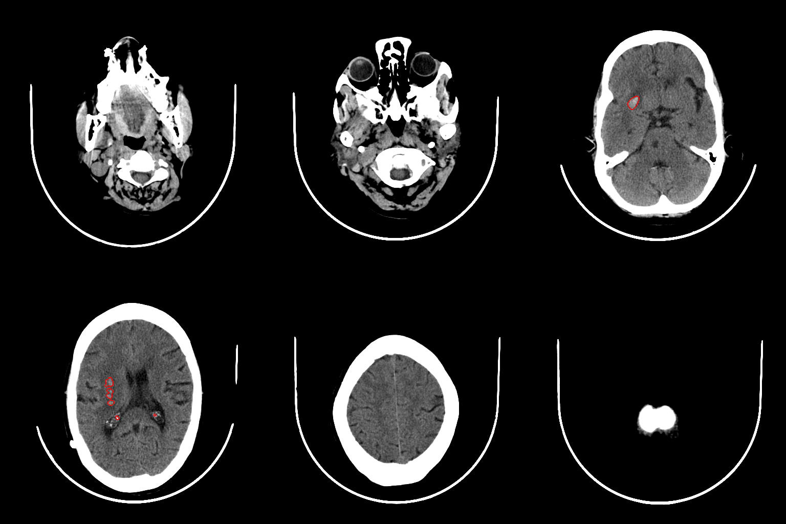
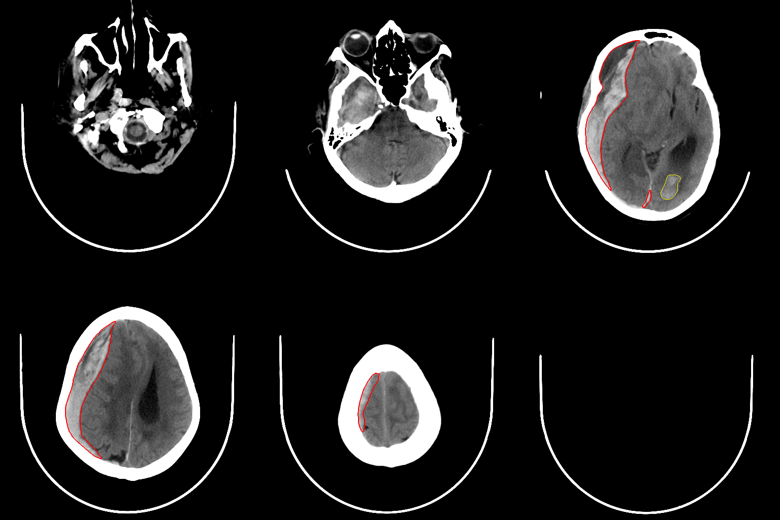
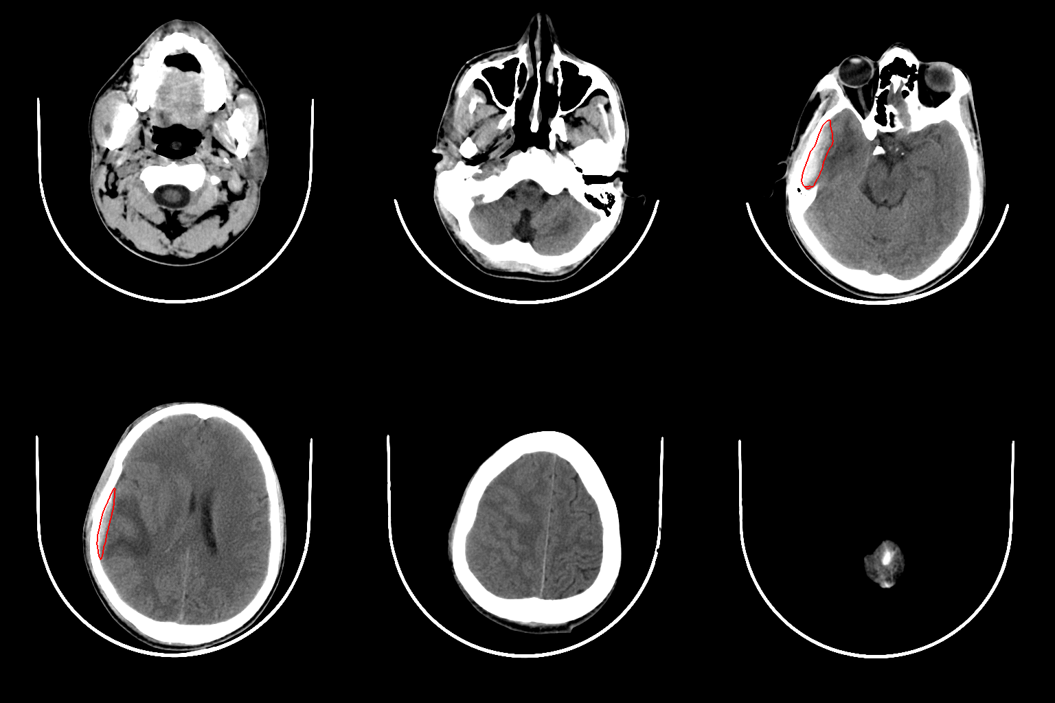
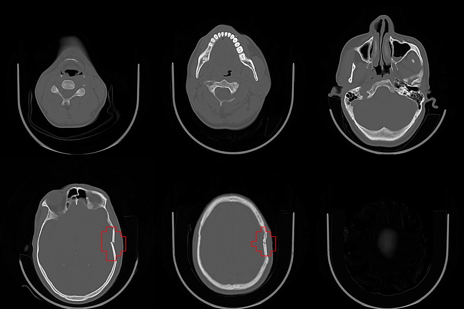
Read our paperNon-contrast head/brain CT is the standard initial imaging study for patients with head trauma or stroke symptoms. In this paper, we describe the development, validation and clinical testing of fully automated deep learning algorithms that are trained to detect abnormalities requiring urgent attention from head CT scans.
We retrospectively collected a dataset containing 313,318 head CT scans along with their clinical reports from various centers. Of these, 21,095 scans (Qure25k dataset) were used to validate and the rest to develop the algorithms.
Additionally, we collected CQ500 dataset from different centers in two batches B1 & B2 to clinically validate the algorithms. CQ500 dataset contained 491 scans.
The gold standard for CQ500 dataset was consensus of three independent radiologists while the clinical report is considered gold standard for Qure25k dataset.
See ROC curvesFinding | CQ500 (95% CI) |
Qure25k (95% CI) |
---|---|---|
Intracranial hemorrhage |
0.9419 (0.9187-0.9651) |
0.9194 (0.9119-0.9269) |
Intraparenchymal | 0.9544 (0.9293-0.9795) |
0.8977 (0.8884-0.9069) |
Intraventricular | 0.9310 (0.8654-0.9965) |
0.9559 (0.9424-0.9694) |
Subdural | 0.9521 (0.9117-0.9925) |
0.9161 (0.9001-0.9321) |
Extradural | 0.9731 (0.9113-1.0000) |
0.9288 (0.9083-0.9494) |
Subarachnoid | 0.9574 (0.9214-0.9934) |
0.9044 (0.8882-0.9205) |
Calvarial fracture | 0.9624 (0.9204-1.0000) |
0.9244 (0.9130-0.9359 |
Midline Shift | 0.9697 (0.9403-0.9991) |
0.9276 (0.9139-0.9413 |
Mass Effect | 0.9216 (0.8883-0.9548) |
0.8583 (0.8462-0.8703) |
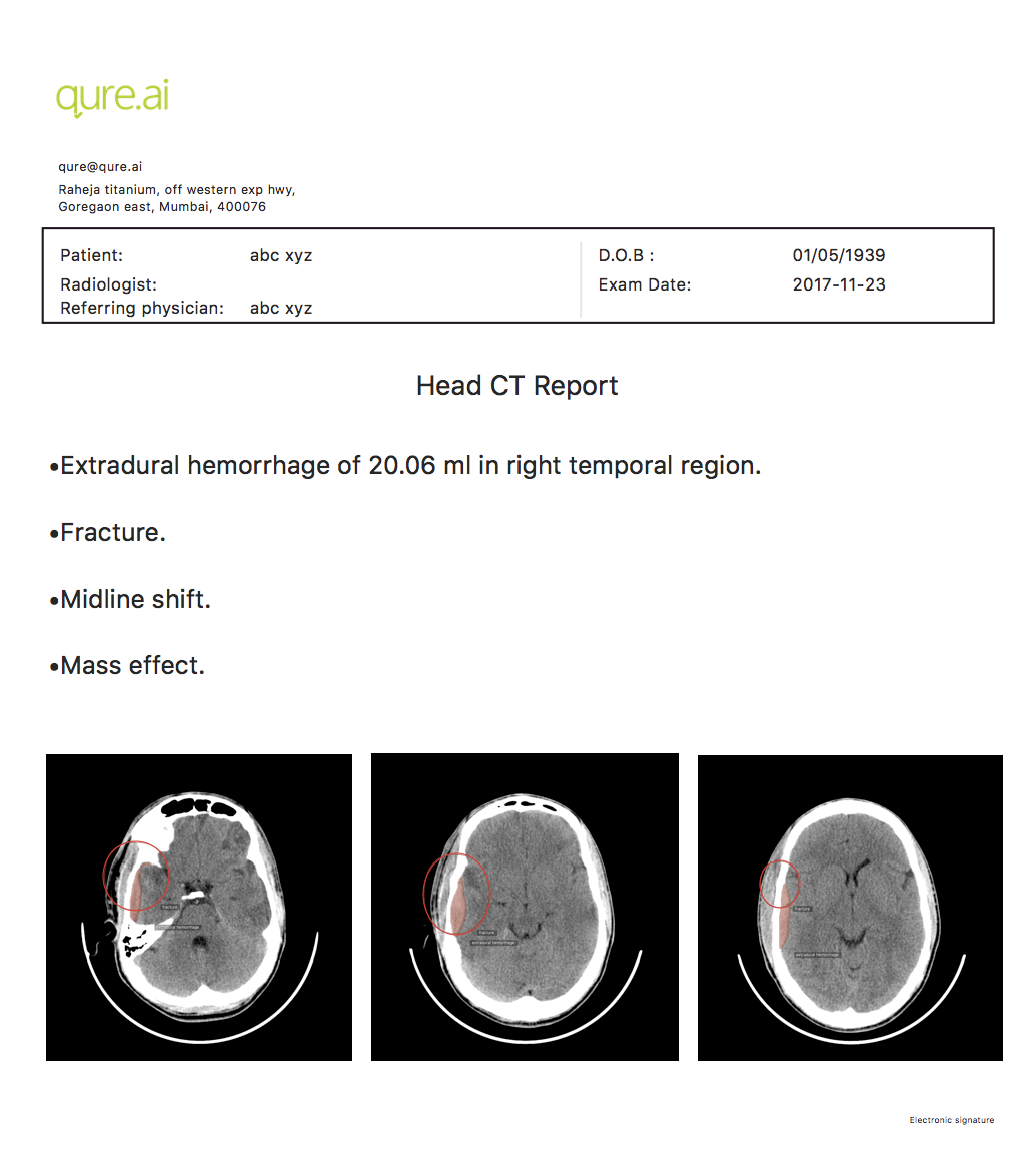
Our algorithms can localize and quantify hemorrhages and fractures. Put together with brain anatomy segmentation algorithms, we can automatically generate reports with anatomical locations of lesions.
We have made the CQ500 dataset of 491 scans with 193,317 slices publicly available so that others can compare and build upon the results we have achieved in the paper
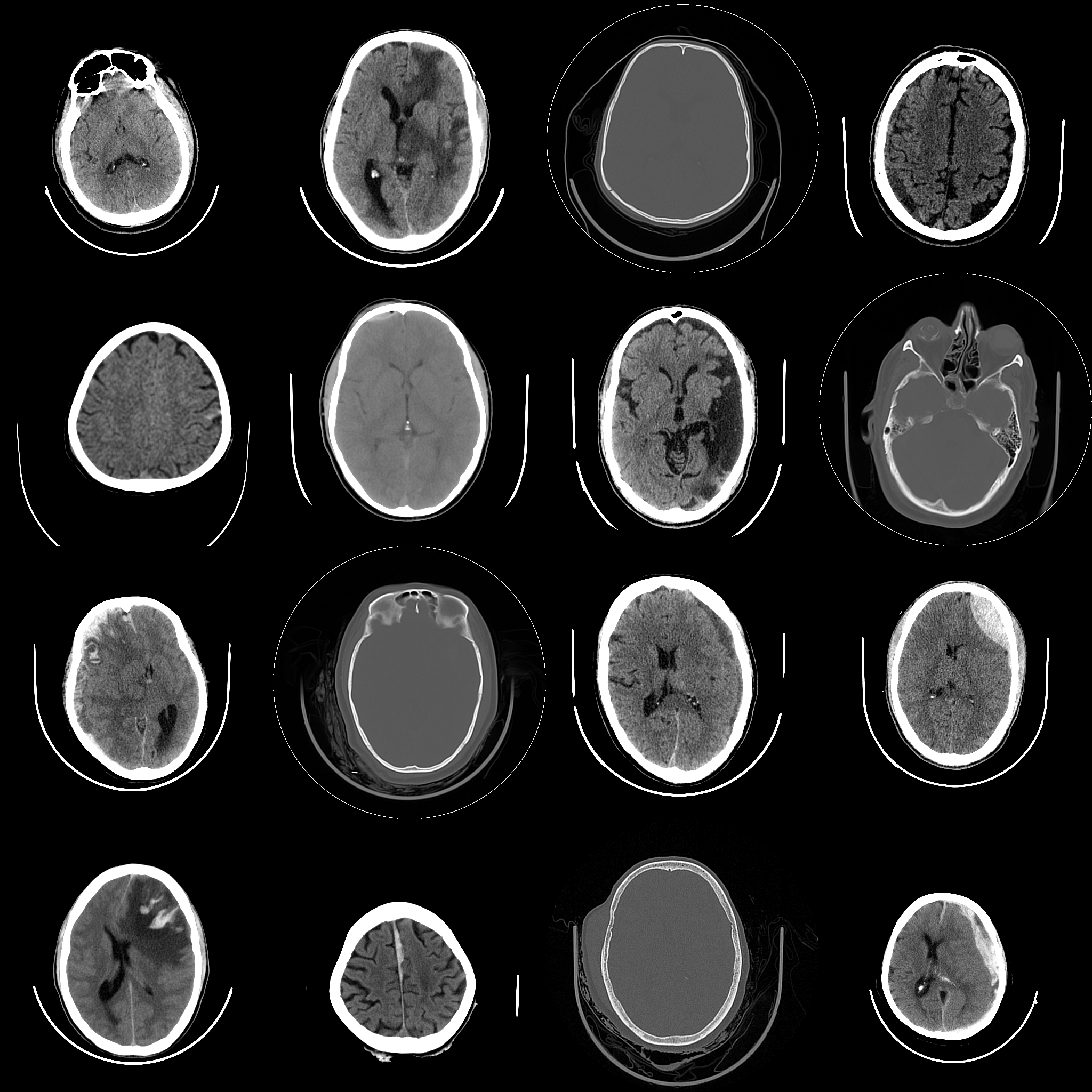
We provide anonymized dicoms for all the scans and the corresponding radiologists' reads. 'Explore data' shows you a sample of this data.
Collaborating with clinicians helps us in carrying further research in head CT scans. Most of our research is done in collaboration with radiologists. If you are interested, please reach out to sasank.chilamkurthy@qure.ai